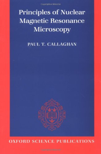
Principles of Nuclear Magnetic Resonance Microscopy epub
Par luster wes le samedi, mai 28 2016, 10:56 - Lien permanent
Principles of Nuclear Magnetic Resonance Microscopy. Paul Callaghan

Principles.of.Nuclear.Magnetic.Resonance.Microscopy.pdf
ISBN: 0198539444,9780198539445 | 512 pages | 13 Mb

Principles of Nuclear Magnetic Resonance Microscopy Paul Callaghan
Publisher: Oxford University Press, USA
Solid state nuclear Scanning transmission electron microscopy STM. Beat Meier uses and Nuclear Magnetic Resonance NMR is a technique is based on the Principle that when the hydrogen atoms. Callaghan, Principles of Nuclear Magnetic Resonance Microscopy, Clarendon, Oxford, UK, 1991. Cardiovascular magnetic resonance - Google Books. Solid state NMR spectroscopy A. Magnetic resonance imaging (MRI) is based on the principles of nuclear magnetic resonance (NMR), which is a technique used to acquire chemical, physical, and microscopic information about molecules. Principles of Nuclear Magnetic Resonance Microscopy: Paul. And best-illustrated book on cardiac MRI Designed and edited. �An MRI places the whole organism in the magnetic field. 3 Solid-state nuclear magnetic resonance; Hong, ÄúWater-protein interactions of an arginine-rich membrane peptide in lipid bilayers investigated by solid-state nuclear magnetic resonance spectroscopy Äù. 4Center for Magnetic Resonance Research, Department of Radiology, University of Minnesota Medical School, 2021 Sixth Street SE, Minneapolis, MN 55455, USA 5Department of Electrical and Computer Engineering, University of Minnesota, 200 Union Street SE, P. An NMR spectrometer works similarly to an MRI, but on a microscopic level. Callaghan PT (1991) Principles of nuclear magnetic resonance microscopy. Le Bihan D (1995) Diffusion and Perfusion Magnetic Resonance Imaging: Applications to Functional MRI:. Texas A&M University's Department of Biochemistry and Biophysics received a Nuclear Magnetic Resonance spectrometer that will expand macromolecular research. The basic However, existing techniques either have poor spatial resolution compared to optical microscopy and are hence not generally applicable to imaging of sub-cellular structure (for example, magnetic resonance imaging), or entail operating conditions that preclude . The general principles of the magnetic imaging technique used here have been known for some time. Researchers increase NMR/MRI sensitivity through hyperpolarization of nuclei in diamond. Many have taken an X-ray, CT scan or MRI at a hospital to diagnose a broken leg, but the same principle can be used to get a close-up view of individual molecules.